
Diagnostic testing is crucial for clarifying diagnoses, staging diseases, monitoring therapy, and assessing complications; proper preparation is key for accurate results.
Modern medicine heavily relies on lab tests, with resources like Mosby’s reference offering guidance on test preparation and performance to ensure safety and precision.
Comprehensive manuals, such as those from Texas DSHS, detail laboratory tests for diseases, directly impacting patient care and overall clinical outcomes significantly.
A. The Role of Lab Tests in Modern Medicine
Lab tests are foundational to contemporary medical practice, serving as indispensable tools for clinicians across diverse specialties. They aren’t merely confirmatory; they actively guide diagnostic pathways, enabling precise identification of ailments and their underlying causes. Resources like the Mosby’s Diagnostic and Laboratory Test Reference emphasize the importance of understanding test nuances to minimize errors and avoid unnecessary repetitions.
Effective patient management hinges on accurate laboratory data. Tests facilitate disease staging, allowing for tailored treatment plans, and are vital for therapeutic monitoring, ensuring interventions are effective and adjusting them as needed. Furthermore, lab results are crucial in assessing potential complications, enabling proactive intervention. The latest research, reflected in resources like the University of Iowa’s guides, continually refines our understanding of test values and their clinical implications.
Ultimately, the judicious ordering and interpretation of lab tests, guided by comprehensive manuals, directly translates to improved patient outcomes and a more efficient healthcare system.
B. Types of Diagnostic Tests: An Overview
Diagnostic testing encompasses a broad spectrum of methodologies, extending far beyond traditional laboratory analyses. Core laboratory tests, like hematology and clinical chemistry, provide fundamental insights into a patient’s physiological state. However, specialized tests, including microbiology, immunology, and increasingly, molecular diagnostics like PCR, offer targeted assessments for specific conditions.
Beyond the laboratory, diagnostic imaging techniques – X-rays, CT scans, MRI, ultrasound, and nuclear medicine – provide visual representations of internal structures and functions. A comprehensive understanding, often detailed in manuals like Mosby’s, is crucial for selecting the appropriate test based on clinical indications and potential contraindications.
Effective test selection considers whether results will genuinely alter patient management, avoiding unnecessary harm or wasted resources. Resources emphasize the importance of addressing potential pre-analytical challenges, such as hemolysis or analyte instability, to ensure reliable results.

II. Pre-Analytical Considerations
Patient preparation and proper specimen collection are vital; guidelines in resources minimize errors, ensuring test accuracy and preventing unnecessary repeats due to issues.
A. Patient Preparation for Testing
Effective patient preparation is paramount for reliable diagnostic results, directly influencing the accuracy of subsequent analysis and clinical decision-making. Comprehensive manuals, like Mosby’s Diagnostic and Laboratory Test Reference, emphasize detailed guidelines tailored to each specific test.
These guidelines often encompass fasting requirements, medication restrictions, and hydration protocols, all designed to minimize variables that could skew results. Understanding contraindications – situations where testing won’t alter management or poses undue harm – is equally crucial.
Proper instruction and clear communication with patients are essential to ensure compliance and reduce the likelihood of sample rejection or the need for re-testing. A well-prepared patient contributes significantly to a streamlined and accurate diagnostic process, ultimately improving patient care outcomes.
B. Specimen Collection Techniques
Accurate specimen collection is a cornerstone of reliable laboratory testing, demanding strict adherence to standardized protocols detailed in diagnostic manuals. These techniques vary significantly depending on the test, encompassing blood draws, urine collection, and tissue biopsies.
Minimizing pre-analytical errors, such as hemolysis or contamination, is critical; proper training and quality control measures are essential for personnel. The “Users Guide to Test Preparation and Performance” within resources like Mosby’s emphasizes meticulous technique.
Correct labeling, storage, and transportation of specimens are equally vital to maintain sample integrity. Following established guidelines ensures the analyte remains stable and representative of the patient’s condition, leading to accurate and clinically relevant results.
C. Common Sources of Error in Pre-Analysis
Pre-analytical errors represent a significant source of inaccuracies in laboratory testing, often stemming from issues before the sample reaches the analyzer; Hemolysis, contamination, and analyte instability are frequently cited challenges, as highlighted in resources detailing diagnostic testing.
Operator-dependent variability, stemming from inconsistent technique during collection or handling, can also introduce errors. Furthermore, discrepancies between established guideline decision limits and local reference intervals pose interpretive difficulties.
Inappropriate patient preparation, such as failing to adhere to fasting requirements, or incorrect tube selection can invalidate results. Diagnostic manuals emphasize meticulous attention to detail and robust quality control to mitigate these risks and ensure reliable data.

III. Core Laboratory Tests
Essential tests include hematology, clinical chemistry (electrolytes, glucose, enzymes), and urinalysis, providing fundamental insights into patient health and disease states.
A. Hematology Tests: Complete Blood Count (CBC) and Beyond
Hematology testing, beginning with the Complete Blood Count (CBC), forms a cornerstone of diagnostic evaluation, assessing red blood cell indices, white blood cell differentials, and platelet counts.
CBC analysis aids in identifying anemias, infections, and bleeding disorders, while further hematological investigations, like peripheral blood smears, provide morphological details.

Accurate specimen collection and handling are paramount, as hemolysis or improper anticoagulation can significantly impact results, leading to misinterpretations and potentially incorrect clinical decisions.
Reference ranges must be considered alongside patient history and clinical presentation; variations exist based on age, sex, and ethnicity, necessitating careful interpretation.
Beyond the CBC, specialized tests like coagulation studies (PT, PTT) and bone marrow aspiration/biopsy offer deeper insights into hematological conditions, guiding targeted therapies.
B. Clinical Chemistry Tests: Electrolytes, Glucose, and Enzymes
Clinical chemistry encompasses a broad spectrum of tests evaluating various biochemical parameters within bodily fluids, primarily blood and urine, offering crucial diagnostic insights.
Electrolyte panels (sodium, potassium, chloride) assess fluid balance and nerve/muscle function, while glucose measurements are vital for diagnosing and monitoring diabetes mellitus.
Enzyme assays (e.g., liver enzymes, amylase, lipase) indicate tissue damage or dysfunction; careful attention to pre-analytical factors is essential for accurate enzyme determination.
Hemolysis, improper storage, or delayed analysis can significantly alter enzyme levels, leading to false results and potentially inappropriate treatment strategies.
Reference intervals, often provided by the laboratory, must be interpreted in the context of the patient’s clinical presentation and any concurrent medications or conditions.
C. Urinalysis: Components and Clinical Significance
Urinalysis is a fundamental diagnostic tool, involving physical, chemical, and microscopic examination of urine, providing valuable clues about kidney function and systemic diseases.
Physical examination assesses color, clarity, and specific gravity, while chemical analysis detects substances like protein, glucose, ketones, blood, and bilirubin.
Microscopic examination identifies cells (red blood cells, white blood cells, epithelial cells), casts, and crystals, aiding in diagnosis of infections or kidney disorders.
Accuracy in urinalysis relies on proper collection techniques and timely analysis, as delaying testing can lead to inaccurate results due to bacterial overgrowth.
Clinical correlation is crucial; abnormal findings must be interpreted alongside the patient’s history, physical exam, and other laboratory data for a comprehensive assessment.

IV. Specialized Laboratory Tests
Specialized tests encompass microbiology for identifying infectious agents, immunology for assessing immune function, and molecular diagnostics like PCR for genetic analysis.
A. Microbiology Tests: Identifying Infectious Agents
Microbiology tests are fundamental in diagnosing infectious diseases, directly influencing patient care and treatment outcomes, particularly within hospital settings. These tests encompass a broad range of techniques designed to identify bacteria, viruses, fungi, and parasites responsible for infections.
Common methods include culturing specimens – such as blood, urine, or tissue – to grow and identify microorganisms. Microscopic examination allows for direct observation of pathogens, while staining techniques enhance visualization. Increasingly, molecular methods like PCR are employed for rapid and highly sensitive detection of microbial DNA or RNA.
Accurate identification is crucial for selecting appropriate antimicrobial therapy and preventing the spread of infection. Laboratories adhere to strict quality control measures to ensure reliable results, impacting clinical decisions and public health initiatives.
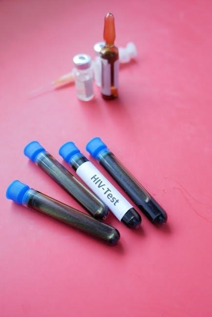
B. Immunology and Serology Tests: Assessing Immune Function
Immunology and serology tests evaluate the function of the immune system and detect the presence of antibodies or antigens, providing insights into a patient’s immune response. These tests are vital in diagnosing autoimmune diseases, immunodeficiencies, and infectious diseases where antibody detection is key.
Common assays include enzyme-linked immunosorbent assays (ELISAs) for quantifying antibodies, flow cytometry for analyzing immune cell populations, and complement fixation tests. These techniques help determine if the body is mounting an appropriate immune response to a pathogen or if the immune system is attacking its own tissues.
Reliable results are essential for guiding treatment decisions, such as immunosuppressive therapy for autoimmune conditions or immunoglobulin replacement therapy for immunodeficiencies, directly impacting patient health.
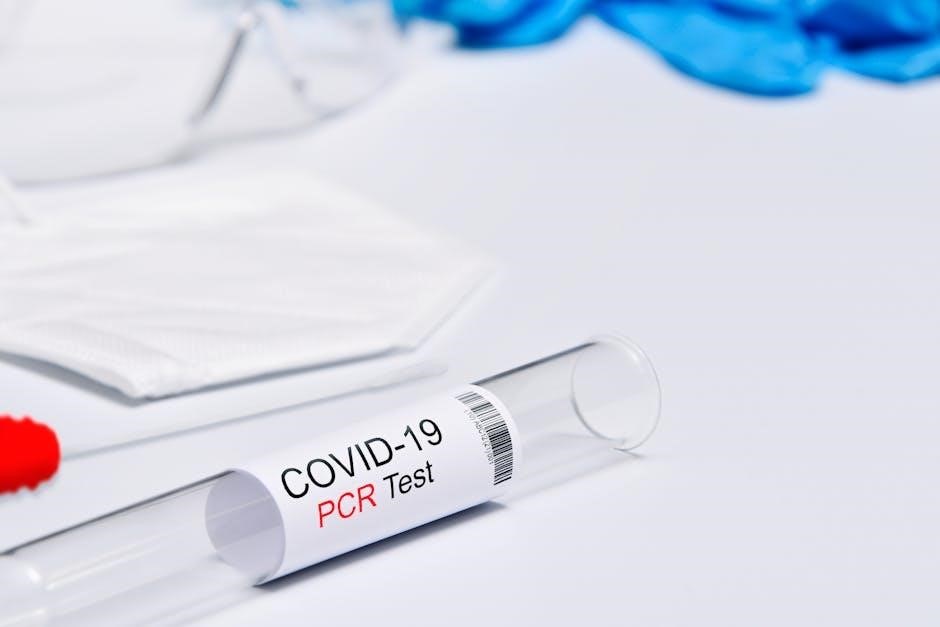
C. Molecular Diagnostics: PCR and Genetic Testing
Molecular diagnostics, encompassing techniques like Polymerase Chain Reaction (PCR) and genetic testing, represent a powerful advancement in disease detection and personalized medicine. PCR amplifies specific DNA sequences, enabling the rapid identification of infectious agents, genetic mutations, and cancer biomarkers with high sensitivity.
Genetic testing analyzes an individual’s DNA to assess their risk for inherited diseases, predict drug response (pharmacogenomics), and diagnose genetic disorders. These tests are increasingly used in oncology to guide targeted therapies based on tumor genetic profiles.
Accurate interpretation of molecular test results requires specialized expertise and careful consideration of potential false positives or negatives, ensuring appropriate clinical management and improved patient outcomes.

V. Diagnostic Imaging Techniques
Imaging modalities, including X-rays, CT scans, MRI, ultrasound, and nuclear medicine, visualize internal structures, aiding diagnosis and monitoring disease progression effectively.
A. X-rays and Computed Tomography (CT) Scans
X-rays utilize electromagnetic radiation to produce images of bones and dense tissues, offering a rapid and cost-effective initial assessment for fractures or foreign objects.
Computed Tomography (CT) scans, however, employ X-rays with computer processing to create detailed cross-sectional images of the body, revealing soft tissues, blood vessels, and subtle bone abnormalities.
Preparation for CT scans often involves contrast dye administration to enhance visualization, though potential allergic reactions or kidney concerns must be considered.
Both techniques are invaluable in diagnosing conditions like pneumonia, internal bleeding, and cancers, but involve radiation exposure, necessitating careful justification and minimization of dose.
Proper positioning and technique are crucial for image quality, and interpretation requires expertise to differentiate normal anatomy from pathological findings accurately.
B. Magnetic Resonance Imaging (MRI)
Magnetic Resonance Imaging (MRI) utilizes strong magnetic fields and radio waves to generate detailed images of organs and tissues without ionizing radiation, offering superior soft tissue contrast.
Preparation for MRI requires careful screening for metallic implants, as the magnetic field can pose risks; patients often receive detailed instructions regarding fasting or medication adjustments.
MRI excels in visualizing the brain, spinal cord, joints, and internal organs, aiding in the diagnosis of neurological disorders, musculoskeletal injuries, and cancers.
Different MRI sequences highlight specific tissue characteristics, allowing for nuanced assessments of inflammation, edema, and structural abnormalities.
The procedure is generally painless but can be lengthy and require patients to remain still within a confined space, potentially causing claustrophobia.
C. Ultrasound and Nuclear Medicine
Ultrasound employs high-frequency sound waves to create real-time images of internal structures, proving invaluable for obstetrics, cardiology, and abdominal assessments; it’s non-invasive and portable.
Nuclear medicine utilizes radioactive tracers to visualize organ function and detect abnormalities at a cellular level, offering unique diagnostic capabilities.
Common nuclear medicine scans include bone scans, cardiac stress tests, and PET scans, aiding in cancer detection, heart disease diagnosis, and neurological evaluations.
Patient preparation varies depending on the scan, often involving fasting or intravenous injection of the tracer; radiation exposure is minimized and carefully monitored.
Image interpretation requires specialized expertise, correlating anatomical findings with physiological function to provide comprehensive diagnostic insights.

VI. Interpreting and Evaluating Test Results
Accurate interpretation demands understanding reference ranges, recognizing potential false positives/negatives, and crucially, correlating lab findings with the patient’s overall clinical presentation.
A. Reference Ranges and Normal Values
Establishing reference ranges is fundamental to interpreting laboratory results, representing the values found in a healthy population. However, these ranges aren’t absolute; variations exist based on age, sex, ethnicity, and even geographic location.
Diagnostic manuals emphasize the importance of understanding that “normal” is a statistical construct, not a definitive boundary. Local laboratories often establish their own reference intervals, potentially differing from guideline decision limits.
Discordance between these local ranges and standardized guidelines can lead to misinterpretation, highlighting the need for clinicians to be familiar with the specific lab’s established values. Careful consideration of these nuances is vital for accurate clinical assessment and avoiding unnecessary interventions based on misinterpreted data.
Furthermore, understanding the statistical methods used to derive these ranges—mean, standard deviation, percentiles—provides a deeper appreciation for their limitations and appropriate application in patient care.
B. Understanding False Positives and False Negatives
Diagnostic testing isn’t infallible; both false positive and false negative results can occur, impacting patient management. A false positive indicates a condition is present when it isn’t, potentially leading to unnecessary anxiety and further, invasive testing.
Conversely, a false negative suggests a condition is absent when it’s actually present, delaying appropriate treatment and potentially worsening outcomes. Pre-analytical errors – hemolysis, contamination, or unstable analytes – significantly contribute to inaccurate results.
Manuals stress the importance of recognizing factors influencing these errors, including operator variability and limitations of the test itself. Clinical correlation is crucial; lab findings should always be interpreted in the context of the patient’s overall clinical picture.
Careful evaluation and repeat testing, when appropriate, can help minimize the impact of these inaccuracies on patient care decisions.
C. Clinical Correlation of Lab Findings
Interpreting laboratory results requires more than simply comparing values to reference ranges; clinical correlation is paramount. A manual of diagnostic tests emphasizes that lab findings must be considered alongside the patient’s complete medical history, physical examination, and presenting symptoms.
Discordance between lab results and clinical presentation should prompt further investigation, not automatic acceptance of the lab value. Factors like patient age, medication use, and comorbidities can influence test results, necessitating careful consideration.
Guideline decision limits and local reference intervals may also vary, requiring awareness of laboratory-specific data. Ultimately, the physician’s judgment, informed by all available information, determines the appropriate course of action.
Effective patient care hinges on integrating lab data with the broader clinical context, avoiding reliance on isolated test results.
VII. Quality Control and Assurance in the Laboratory
Maintaining accuracy and reliability in diagnostic testing demands rigorous quality control and assurance protocols. A comprehensive manual of diagnostic and laboratory tests details these essential procedures, emphasizing minimizing errors throughout the testing process.
Pre-analytical errors, such as hemolysis or contamination, must be proactively addressed through standardized specimen collection and handling techniques. Internal quality control involves regular analysis of known samples to monitor instrument performance and reagent stability.
External proficiency testing, where labs analyze unknown samples from an external source, validates accuracy against peer institutions. Continuous monitoring and documentation of all quality control data are crucial for identifying and rectifying any deviations from established standards.
Adherence to these principles ensures dependable test results, ultimately benefiting patient care.